① ‘IFSCC 2023’ 리뷰 〈4〉

2023년 9월 4~7일 스페인 바르셀로나에서 열린 ‘제33회 IFSCC Congress’ 리뷰를 이번호로 마무리한다. 피부 기반기술 개발 사업단(단장 황재성, 경희대 유전생명공학과 교수)과 더케이뷰티사이언스가 공동으로 진행했다. _ 편집자 주 


Rethinking Science - Understanding skin sensoriality and function

① ‘IFSCC 2023’ 리뷰 <4>

신진희 P&K피부임상연구센타 중앙연구소, 이사
연구 동향 
Keynote - ‘홀리스틱 뷰티(Holistic Beauty)’

홀리스틱 뷰티(Holistic Beauty)는 피부의 단편적 아름다움 뿐 아니라 몸 전체 시스템과 바이탈(Vital), 멘탈(mental)까지 아우르면서 연결되어 진정한 아름다움이 표현되는 것을 의미한다. 홀리스틱 뷰티는 혈관계, 면역계, 신경계를 통해서 인식될 수 있다. 일반적으로 혈관이 피하지방층 아래에 존재하고 피부에는 없는 것으로 인식하고 있지만, 노화가 되면서 미세혈관은 감소하고 불규칙한 모양으로 변화한다. 이러한 미세혈관의 변화는 피부탄력과 상관관계가 높아 미세혈관이 피부 탄력 유지의 지지대 역할을 할 수 있다고 본다. 미세혈관의 감소는 피부 진피층 구성단백질 생성에 필요한 성분을 공급하지 못하므로 노화가 되면 콜라겐 단백질 생성이 연쇄적으로 감소 된다고 역학적으로 설명할 수 있다. 특히 표피까지 뻗어나온 혈관은 표피의 줄기세포에 소스를 공급하는데 ‘Netrin-1’을 통해 미세혈관과 표피 사이의 적당한 거리를 유지하도록 한다. 면역계는 생체리듬과 밀접하게 연관되어 있어 수면패턴의 문제는 피부 건강에 필요한 면역계 요소들의 혼란을 야기할 수 있다. 촉각적 터치와 감성적 터치는 피부 내 ‘Merkel cell’과 신경섬유와 교접하며 오감은 신체와 신경을 통해 소통하는 것이다. 즉, 신경세포가 콜라겐 합성에도 연관되어 있다는 것을 생체외시스템을 통해 확인할 수 있다. 앞으로 홀리스틱 뷰티를 통한 피부 아름다움의 이해는 세포에서부터 각 조직에서 몸 전체 시스템에 이르는 통합적인 이해와 솔루션이 필요할 수도 있다. 
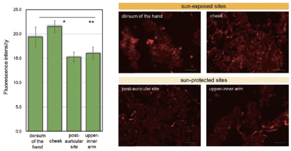
그림 1. Representative images of 3-NT visualized in corneocytes by immunostaining: differences of sunprotectedand sun-exposed skin sites. Corneocytes were collected with tape-stripping by cellotape(Nichiban, Tokyo), and were transferred to glass slides by immersion into xylene twice. Corneocytes were immuno-stained with a primary 3-N antibody(diluted 1:200) and Secondary and antibody, anti-rabbit lgG conjugated AlexaFluor594(diluted 1:200). Fluorescence images of corneocytes were obtained using a BZ-X810 All-in-One fluorescence microscope. Scale bars: 100um. The FI originating from 3-NT in fluorescence images was quantified using specialized software, corneocytometry 2. Results are presented as means ±SD, n3. Dunnett test, *p 〈0.05, ** p〈 0.01
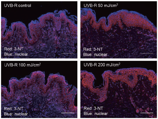
그림 2. Representative images of 3-NT visualized inUVBirradiated human excised skin. Excised human skin was exposed to UVB(0, 50, 100 or 200mJ/cm²) and was further cultured for 3 days after which thin sections were immuno-stained with the 3-NT antibody. Nuclei were stained with Hoechst 33342. Scale bars: 100um. red:3-NT, blue:nuclei


더 자세한 내용은 네이버 화장품과학'(유료)에서 읽을 수 있습니다.

 
저작권자 © THE K BEAUTY SCIENCE 무단전재 및 재배포 금지